Novel Gene Therapies to Treat Usher Syndrome Type 1F: Mini-PCDH15
In a second strategy to circumvent the size limitation of PCDH15, we used the exquisite atomic structures of PCDH15 solved by Marcos Sotomayor and others to identify domains that may not be essential for function and that might be removed. PCDH15 has 11 extracellular cadherin (EC) repeats that act like links in a chain. EC1-3 bind the tip-link protein CDH23, EC11 and the following MAD12 domain may interact with the TMC1 transduction channel, but the intervening seven EC repeats might be dispensable. Using the atomic structure, we designed eight mini-PCDH15 versions in which the Ca2+-binding residues of the EC-EC junctions were preserved. Three of these were short enough to be encoded in single AAV vectors and were tested in the inner ears of a mouse model of Usher 1F. One of them almost completely prevents the hair cell degeneration and hearing loss in this mouse model, and it prevents the vestibular deficit in the mouse. | |
| |
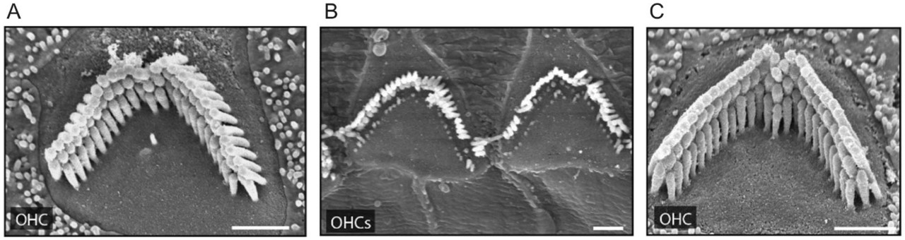
Preservation of hair cells in a mouse cochlea with mini-PCDH15 gene therapy. A, The normal bundle of sensory cilia on one hair cell at 30 days of age. B, Two hair cells from an untreated Usher 1F mouse showing extensive degeneration by 30 days. The mice are deaf. C, A hair cell from an Usher 1F mouse that received the mini-PCDH15 therapy at birth. The bundle looks healthy and the mice respond to sound. | |
It may be more feasible to prevent the slow loss of vision in Usher 1F patients than to prevent the congenital deafness. Mice lacking PCDH15 have little or no retinal phenotype, so we are exploring another animal model of Usher 1F, a pcdh15b knockout zebrafish developed by Jen Phillips and Monte Westerfield. These zebrafish show degeneration of photoreceptors and impaired visual function in 7-day larvae, so they are an excellent platform for rapid testing of therapies. We are testing mini-PCDH15B versions by transposon insertion into the zebrafish genomes, using both immunogold EM for assessing proper localization in photoreceptors and physiological tests for assessing retinal function. | |
| |
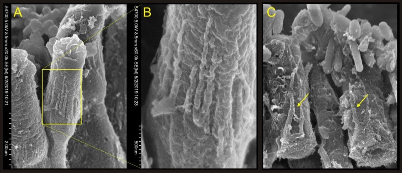
Pathology of photoreceptors in pcdh15b knockout zebrafish. A, Photoreceptors in a 7 day wild-type control zebrafish, in the region of the outer segments. B, Inset showing the palisade-like calyceal process that are associated with PCDH15. C, Outer segments from a pcdh15b mutant zebrafish. The outer segments of the photoreceptors are disorganized and the calyceal process are mis-formed. | |
This project is a collaboration with the laboratories of Artur Indzhykulian, Marcos Sotomayor and Monte Westerfield. |